An Adventure
in Protozoan Art
Come on in, the water's fine!! The following pics were taken through my microscope using a digital camera. Specimens were from my backyard pond.
____________ * * * * * * * * * * * * * * * ____________
Paramecium caudatum, an image of one of the more common Paramecium types and one of the smaller varieties, growing to about 125 microns in size.
Paramecium bursaria, a species of Paramecium that appears green because of the presence of symbiotic Zoochlorella.
This image shows in unusual color the intricate details of this amazing one celled animal - the Paramecium.
Click here to see an image of a Paramecium dividing.
Paramecia have a defense system that they can deploy when attacked or when they feel threatened. Click here to find out more.
All ciliates also can multiply by a process known as conjugation. Here is a rare image of two Paramecium engaged in true sexual reproduction.
Get a real sense of the size of these critters. Click on this link to see a swarm of Paramecium inside a speck of water that is smaller than the "period" on your keyboard's "period key".
Actinosphaerium (or an Actinophyrs). The spikes are used to spear other protozoa. The protoplasmic "soup" is then sucked out of the victim into a food vacuole where it is digested.
An excellent image of an Actinophyrs digesting a meal.
An excellent image of an Actinosphaerium. Actinosphaerium can grow up to 1,000 microns in size. This specimen certainly is close to that figure.
An image of three different Protozoa, all in a space not much bigger than a pin head.
Vorticella. Cilia create currents in the water to draw food into the Vorticella's huge anterior opening. Bits of ingested food are clearly visible. Because the cilia are constantly moving, they are blurred in the photo but still visible.
There are in excess of 100 species of Vorticella. Here is an image of a species that clearly shows all of the creature's internal parts and functions.
Another image of a Vorticella. The trailing stalk is faintly visible in this photo. See the picture along with a description of the Vorticella's unusual behavior.
I had to follow these five Vorticella around for several minutes before they settled in one spot. Click here to see what happens when they're startled.
Click on this link to see a bit of misfortune that befell an inattentive Vorticella.
Here is an image of a Vorticella dividing.
Here's a protozoa that appears quite different from a Vorticella but has many of the same behavior and feeding patterns, the Stentor.
This is an excellent image of two Stentor. Look here.
The Stentor is not a communal creature, but it is possible to encourage them to "huddle" together. Click on the link.
The Stentor provides many opportunities for capturing some interesting images. Here's a Stentor consuming a single celled Closterium.
Here is an image of two Stentor that you Must See.
Here is an image of a blue Stentor showing the large, fully extended mouth opening and the surrounding cilia. Click Here.
I continue to find interesting specimens of the Blue Stentor. Here is a composite image.
This one celled Protozoan continues to show its many fascinating faces. Two more images of the Blue Stentor.
Crustaceans are abundant in ponds and are easily spotted once the water warms. The microscopic ones feed on algae, decaying matter, other protozoa and even bacteria.
Here's a picture of a common crustacean - a Chydorus, a relative of the Daphnia. A dead Chydorus,
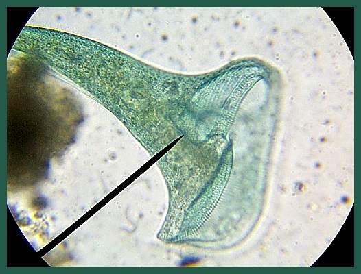
Here's an image of a copepod that doesn't look like a copepod, nor is it free living. It's a parasitic Anchor Worm.
Here is another species of Anchor Worm, commonly called a skin fluke or gill fluke.
When looking for protozoa one wouldn't expect to see anything colorful, since 99% of these microbes are transparent. Click on this link for a pic of a red-colored, and not often seen Blepharisma.
Another picture of a Blepharisma. Click here for a better view of its natural shape and a few of its more prominent features.
The life of a microbe in a pond isn't all fun and games, or eat and sleep. Click here to see how bad things can be.
Here is common pond creature - a Holotrich, and most likely a Dileptus species.
A drop of water from our birdbath contains an assortment of different microbes.
A water drop teeming with micro life, some as small as 1 or 2 microns. This culture was made by allowing dried leaves and grass to decay in the open air.
An Actinosphaerium and a Colurella together. I watched the two of them bump into each other for several minutes and neither one seemed to be bothered by the other. Apparently they aren't part of each other's diet.
Any pond or pond filter will contain a variety of worms. Many of these wrigglers are the larvae of the many different kinds of flies and midges that hang around backyard ponds and ornamental waterfalls, while some are true aquatic worms. A few are visible, such as the bloodworms that we frequently see in our filter systems. Many, however, are microscopic. Click here to see the one worm that must be one of the ugliest and most repulsive in all of wormdom.
Here's an image of a fly larva that takes up residence in the busiest part of the pond. Click here to see why and how.
Glassworms, Bloodworms and Ghostworms are all the larvae of the many different kinds of black flies (midges) that we see around our ponds. Click here to see a Bloodworm and an image of the pupa.
This image is a sequel to the preceeding image. Click here to see the scavengers that have moved in on the remains.
Amoebas are amorphous so they are seen in a variety of shapes and sizes. They also have developed a variety of ways to cope with the dangers of their environment. Click here to see an image of an Arcella, an Amoeba that has developed an unusual method of protection.
Here is another species of Shelled Amoeba. Unlike the Arcella in the previous image, this species clearly shows the gritty, granular composition of the domed shell.
Here is a composite picture of two Shelled Amoebas, both taken from the same water drop. Note the differences in the shell composition, and the evidence of some structural failure on one of them. Click here.
Click here to see an Amoeba the way we always imagined one would look - like an amorphous, protoplasmic "splat".
Here are two images of another naked amoeba, probably an Amoeba proteus, taken just a few seconds apart.
Here is a closer view of the same Amoeba that appears in the previous image.
A common species of Rotifer. Rotifers can be found wherever there is water or moisture - ponds, puddles, damp ground or mosses. They are included in the small-sized group of pond critters, but are easily seen even under low power. There are many varieties and their varied methods of locomotion are interesting to observe. They are usually found on the pond bottom.
Another good place to look for Rotifers is the outdoor bird bath, preferably one that hasn't been cleaned in a week or so. Click here to see a colony of them.
Here is an unusually detailed and clear image of another species of Rotifer. Click here.
Water Mites can be difficult to identify because they are so abundant and varied in appearance. This critter was originally mis-identified as a water mite but in fact is a Cyclops, quite common as both a marine and freshwater species. Click to find out more about these interesting creatures.
Click here for an image of a true water Water Mite, and some additional information about microanimals.
Gastrotrichs are frequent vistors to any backyard pond. They, too, are members of the multicellular Animal Kingdom.
Here is a pic of a microbe from a group of creatures that includes at least four families, thirty genera and numerous species.
Have a look.
Here is an image of what appears to be algae, but really isn't. I included it here because it is readily known by its common name - Blue/Green Algae.
Here is another image of a Blue/Green Algae - likewise, not an algae but one of the many species of Cyanobacteria.
Here are two excellent images of String Algae.
This Algae can cause serious problems if you don't pay attention to your maintenance duties.
Closterium. An interesting Green Protist - easy to spot and quite common in healthy ponds and lakes.
Selenastrum and Pediastrium, two interesting Green Protists. Common in quiet waters, and sometimes found in wet soils and sands.
An unusually clear image of two clusters of Pediastrium.
Some large clusters of Selenastrum. Pediastrium and Selenastrum are just two of the many single cell colonial Protists that give us our dreaded green pond water.
A scraping from one of my lily stems yielded an assortment of different Algae. More than I can identify.
Here is an image of a cluster of Diatoms. They aren't green protists but I've included them here because they are often found in areas where algae is also found.
This image is from the same salt water tank showing several other types of Diatoms. This image was taken at the same magnification as the previous one so the relative sizes can be compared. These were squeezed from the sponge filter. Note the perfect symmetry of the specimens as well as the interesting coloration of the Diatom in the upper right of the image.
This creature, for the moment, is unidentified. It's about 1,000 microns in size, and I found dozens in a container of pond water that I allowed to become stagnant and polluted.
This specimen is large, at least 1,000 microns across, perhaps larger. It was an "unidentified" for quite some time until I was sent a photograph of its twin brother by someone visiting my site. It's a Bryozoa. There is an extensive fossil record of these multicelled critters. They are found in both fresh and sea water and they feed through cilia that cover their bodies. It's an interesting specimen under the microscope.
A Few Words About Size
Interesting Facts and Trivia that Everyone Should Know
Specimen Backgrounds Courtesy of Wim von Egmond. Click Here for Wim's Marvelous Site
Click Here For Another Source For Quality Microscopes, Australia Based
Click Here For - Pond Critters, An Illustrated Guide
Ron DeAngelis - Pond Scum Developer, Owner


Microscopes HQ Everything Microscopes, from About Microscopes to Zoom Microscope
Meet The Silly Cells A Fun Educational Site